Welcome to GS-CMLS
The Graduate School for Computing in Medicine and Life Sciences was established in 2007 within the framework of the Excellence Initiative of the German federal and state governments.
It aims to integrate medicine, computer science and life sciences through structured international doctoral education rooted in a strong research foundation.

Doctoral Programme
The mission of the Graduate School for Computing in Medicine and Life Sciences is to train outstanding doctoral students through a structured teaching and research programme within an interdisciplinary and international environment. The programme for doctoral education is characterized by professional supervision and a quality curriculum.
Having successfully passed the doctoral programme you can receive permission to graduate. The following degrees are awarded by the sections Natural Sciences and Computer Science/ Engineering: Dr. rer. nat. and Dr.-Ing.
Supervision
In order to enrich the scientific and personal development of the young researchers, each student admitted to the doctoral programme is assigned a supervision group consisting of two supervisors and an advisor.
The supervision group gives personal guidance and support to the student with regard to the research project as well as the doctorate in general. Meetings are held on a regularly basis. A progress report is provided to assist the documentation process of the meeting and to keep a record of the student's activity.
Graduate School for Computing in Medicine & Life Sciences
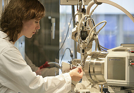
The Graduate School for Computing in Medicine and Life Sciences (GS-CMLS) was established in 2007 in the framework of the Excellence Initiative of the German Governments. It links the three main fields of research of the university: medicine, computer science and life sciences. All main facilities are located on one campus. Around 60 doctoral students from 15 countries are currently enrolled. In the past 10 years more than 50 doctoral students have graduated from the Grad School embarking on successful careers in academia and industry. The Grad School is now a leading research centre in several fields e.g. medical robotics, optical coherence tomography and magnetic particle imaging.
Information for prospective doctoral candidates
Prospective doctoral candidates are advised to contact potential supervisors for research projects they are interested in. Here you can find a list of associated institutes.
News & Events
Neuroscience Lecture WS19/20
by Prof. Lisa Marshall, Institute of Pharmacology and Toxicology